Intraoperative Spinal Navigation

Intraoperative Spine Surgery Navigation Utilizing LoopX Imaging


Revolutionizing Spine Surgery through Accuracy, Safety, and Minimally Invasive Techniques

Intraoperative spine surgery navigation, combined with LoopX imaging, has revolutionised the field of spine surgery.

This technology has significantly improved patient outcomes by offering enhanced precision, improved safety, and minimally invasive techniques.

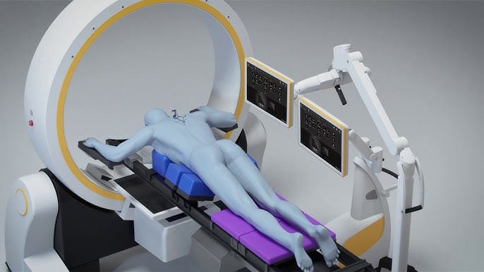
How Navigation with Intraoperative LoopX Imaging Works

Intraoperative navigation, also known as computer-assisted or image-guided surgery, is a technology that enables surgeons to visualise the surgical site in real time using 3D imaging.

During surgery, LoopX imaging, a mobile intraoperative imaging system, acquires high-resolution 3D CT images of the patient's spine. These images are then integrated with the navigation system, allowing the surgeon to view the surgical field with extreme accuracy.

As the surgeon navigates their instruments and implants, such as pedicle screws and inter-body cages, during the procedure, the navigation system tracks the instruments' and implants position. It displays their precise location on the 3D CT images. This real-time guidance helps the surgeon avoid critical structures, ensuring a safer and more accurate surgery.

Essentially the surgeon can see clearly a virtual image of each screw and cage being inserted on a 3D CT scan model on screen as the screw is inserted into the spine. Furthermore, prior to implant insertion the final desired trajectory and position can be planned for a really precise and desirable final implant position.

History of Surgical Navigation and Intraoperative Spinal Imaging

The development of surgical navigation and intraoperative spinal imaging has evolved over several decades.

Early techniques involved using plain X-ray films, which provided limited visualisation of the surgical field. The introduction of fluoroscopy improved visualisation but exposed the patient and surgical team to continuous radiation.

LoopX imaging, introduced in 2019, revolutionised intraoperative spinal imaging by providing high-quality, 3D CT images with reduced radiation exposure. When combined with navigation, this technology significantly improved the accuracy and safety of spine surgery.

Benefits of Intraoperative Spine Surgery Navigation with LoopX Imaging

Intraoperative spine surgery navigation with LoopX imaging has been a major advancement in the field of spine surgery.

This technology has made surgeries safer and more precise by providing real-time, accurate guidance during procedures.

Additionally, it has facilitated the development of minimally invasive techniques, which has improved patient outcomes and shortened recovery times.

As spine surgery continues to evolve, intraoperative navigation and LoopX imaging will remain essential tools for ensuring the best possible patient care.

The development of intraoperative spine surgery navigation utilising LoopX imaging has significantly impacted the field of spine surgery, bringing numerous benefits for patients and surgeons.

As technology advances and more sophisticated systems are introduced, the safety, accuracy, and efficiency of spine surgery are expected to continue improving.

Ultimately, these advances will translate into better patient outcomes, faster recovery times, and an enhanced quality of life for those who undergo spinal surgery.

Minimally Invasive Tubular Lumbar Discectomy

Minimally Invasive Tubular Lumbar Discectomy: A Superior Alternative to Traditional Open Surgery

Minimally invasive tubular lumbar discectomy offers a less invasive approach to treating lumbar disc herniation compared to traditional open surgical discectomy. This innovative technique utilizes a tubular retractor system that enables surgeons to access the spine with minimal disruption to the surrounding tissues, resulting in reduced postoperative pain and faster recovery times.

Indications

Minimally invasive tubular lumbar discectomy is indicated for patients with lumbar disc herniation causing radiculopathy or nerve compression who have not responded to conservative treatments, such as physical therapy, medications, or epidural injections.

Technique

The minimally invasive tubular lumbar discectomy procedure begins with the patient lying face down on the operating table. A small incision is made on one side of the spine, through which a series of dilators are inserted to create a working channel. A tubular retractor is then placed over the dilators to maintain the channel and provide access to the affected area.

Using specialized instruments and working under a microscope, the surgeon removes the portion of the intervertebral disc that is compressing the spinal nerves. Once the nerve compression is relieved, the tubular retractor is removed, and the incision is closed with sutures or staples.

Benefits

  1. Reduced tissue damage: The tubular retractor system minimizes disruption to the surrounding muscles and soft tissues, resulting in less postoperative pain and faster recovery times compared to traditional open surgery.
  2. Smaller incisions: Minimally invasive tubular lumbar discectomy requires only a small incision, leading to reduced scarring and a lower risk of infection.
  3. Lower risk of complications: Due to the minimally invasive nature of the procedure, there is a lower risk of complications such as blood loss, infection, and muscle damage compared to traditional open surgery.
  4. Shorter hospital stays: Patients who undergo minimally invasive tubular lumbar discectomy typically have shorter hospital stays and faster return to normal activities.
  5. Preservation of spinal stability: By accessing the spine through a small incision and avoiding extensive dissection of the surrounding tissues, the procedure preserves the structural integrity of the spine and reduces the risk of spinal instability.

Risks

As with any surgical procedure, there are potential risks associated with minimally invasive tubular lumbar discectomy. These may include infection, bleeding, nerve damage, dural tears, or recurrent disc herniation. However, the overall risk profile is generally considered lower than that of traditional open surgery.

Conclusion

Minimally invasive tubular lumbar discectomy offers a less invasive and more efficient approach to treating lumbar disc herniation. By utilizing a tubular retractor system and performing the discectomy through a small incision, this procedure minimizes tissue damage and results in shorter recovery times compared to traditional open surgery. With its numerous benefits and reduced risk profile, minimally invasive tubular lumbar discectomy is becoming an increasingly popular option for patients seeking relief from debilitating spinal conditions. As technology and surgical techniques continue to advance, this innovative approach is expected to become the gold standard in the treatment of lumbar disc herniation.

Minimally Invasive Lumbar Posterior Interbody Fusion

Minimally Invasive Lumbar Posterior Interbody Fusion: Revolutionizing Spinal Fusion Surgery

Minimally invasive lumbar posterior interbody fusion (MI-RLF) is a cutting-edge surgical technique that offers significant advantages over traditional open and navigated minimally invasive interbody fusion techniques.

This advanced procedure uses sophisticated imaging technology to achieve greater precision, improve patient outcomes, and reduce recovery times.

In this article, we will explore the various interbody fixation options, indications, techniques, benefits, and risks of MI-RLF and discuss the advantages of expanding interbody cages compared to traditional options.

Interbody Fixation Options

Several interbody fusion techniques are available, each with its unique approach to accessing the spine and stabilising the affected vertebrae. These techniques include:

Indications

MI-RLF is indicated for patients with a variety of spinal conditions causing chronic pain and instability, including degenerative disc disease, spondylolisthesis, spinal stenosis, and recurrent disc herniations.

This procedure is recommended for patients who have not found relief through conservative treatments and may be especially beneficial for those who require multi-level fusions or have previously undergone spinal surgery.

Technique

MI-RLF utilises assistance to provide precise, real-time guidance throughout the surgical procedure. After the patient is placed under general anaesthesia, the surgeon makes a small incision in the back and inserts a tubular retractor to create a working channel for the surgical instruments. 

The advanced imaging system helps the surgeon visualise the spine and accurately remove the damaged disc, prepare the endplates for fusion, and insert an interbody cage filled with bone graft material.

Benefits of MI-RLF

Expanding Interbody Cages: A Game-Changer

Expanding interbody cages represent a significant advancement in spinal fusion surgery.

These innovative devices can be adjusted to fit the patient's unique anatomy, providing optimal spinal support and promoting fusion. The benefits of expanding interbody cages include:

Risks

As with any surgical procedure, MI-RLF carries potential risks, including infection, bleeding, nerve injury, and nonunion or malunion of the fused vertebrae. However, the minimally invasive nature of MI-RLF and the use of assistance aims to reduce the likelihood of these complications.

Minimally invasive lumbar posterior interbody fusion represents a major advancement in spinal fusion surgery. By harnessing the power of assistance and advanced imaging technology, MI-RLF offers increased precision, reduced tissue damage, and improved patient outcomes compared to traditional open and navigated minimally invasive techniques.

Patients suffering from chronic back pain and spinal instability should consider consulting with a spine surgeon experienced in MI-RLF to discuss their options and determine if this revolutionary procedure is right for them.

As spinal surgery continues to evolve and incorporate new technologies, the future of minimally invasive spine surgery looks promising. It would offer patients a safer and more effective path to pain relief and restored function.

Anterior Lumbar Interbody Fusion

Anterior Lumbar Interbody Fusion: A Minimally Invasive Approach to Spinal Stability

Anterior lumbar interbody fusion (ALIF) is a minimally invasive surgical technique that aims to treat degenerative spinal conditions, such as disc degeneration, spinal stenosis, and spondylolisthesis.

By approaching the spine from the front, ALIF avoids disrupting back muscles and offers several advantages over traditional posterior interbody fusion techniques.

This article will explore the indications, techniques, benefits, and risks of ALIF, as well as discuss the potential benefits of additional percutaneous pedicle screw fixation.

Indications

ALIF is typically indicated for patients suffering from degenerative spinal conditions causing chronic low back and leg pain that have not responded to conservative treatments.

This procedure may be recommended for those with conditions such as degenerative disc disease, spinal stenosis, or spondylolisthesis. It is also an option for patients who have undergone previous spine surgery and require a revision procedure.

Technique

During an ALIF procedure, the patient is placed under general anaesthesia and positioned on their back. The surgeon makes an incision in the lower abdomen and carefully moves the abdominal muscles and blood vessels aside to access the front of the spine.

The affected disc is removed, and an interbody cage filled with bone graft material is inserted in the disc space to restore the normal height of the vertebral segment.

Once the interbody cage is in place, the surgeon may opt to perform additional percutaneous pedicle screw fixation to enhance spinal stability. This involves the insertion of screws and rods through small incisions in the back, guided by spinal navigation for increased accuracy and precision.

Benefits

  • Minimally invasive: ALIF is performed through a small incision in the abdomen, avoiding the need to disrupt the muscles and soft tissues of the back. Compared to posterior approaches, this results in less postoperative pain and a quicker recovery.
  • Preservation of spinal muscles: Since ALIF does not involve cutting or retracting the back muscles, it preserves their function and reduces the risk of postoperative muscle atrophy and pain.
  • Better access to the disc space: Approaching the spine from the front allows the surgeon to access the affected disc more directly, resulting in a more complete disc removal and more precise placement of the interbody cage.
  • Reduced risk of nerve damage: ALIF reduces the risk of nerve damage by avoiding manipulation of the spinal nerves during surgery.
  • Enhanced spinal stability with pedicle screw fixation: The addition of percutaneous pedicle screw fixation can further stabilise the spine and promote successful fusion without the need for more invasive techniques.

Risks

As with any surgical procedure, ALIF carries potential risks, including infection, bleeding, blood vessel or nerve injury (this technique is sometimes avoided in young men for this reason), and nonunion or pseudarthrosis (failure of the vertebrae to fuse).

However, the overall risk profile is generally lower than traditional open posterior fusion techniques.

Anterior lumbar interbody fusion (ALIF) is a minimally invasive approach to spinal stabilisation that provides several advantages over traditional posterior techniques. With its reduced disruption to spinal muscles and lower risk of nerve damage, ALIF is becoming an increasingly popular choice for patients seeking relief from chronic lower back pain caused by degenerative spinal conditions.

The addition of percutaneous pedicle screw fixation further enhances spinal stability and promotes successful fusion. As surgical technologies and techniques continue to advance, ALIF is expected to remain at the forefront of spine surgery, offering patients a safer and more effective treatment option for debilitating spinal conditions.


Minimally Invasive Tubular Decompression

Minimally Invasive Tubular Lumbar Decompression: A Less Invasive Approach with Improved Outcomes

Minimally invasive tubular lumbar decompression offers a less invasive alternative to traditional open surgical decompression for patients suffering from spinal stenosis or degenerative disc disease.

This advanced procedure utilises a tubular retractor system to access the spine, resulting in reduced tissue damage and faster recovery times.

In this article, we will discuss the indications, techniques, benefits, and risks of this procedure and highlight its advantages compared to traditional open surgery.

Indications

Minimally invasive tubular lumbar decompression is indicated for patients with lumbar spinal stenosis or degenerative disc disease who have not responded to conservative treatments, such as physical therapy, medications, or epidural injections.

This procedure is particularly suitable for patients who require decompression but wish to avoid the risks and longer recovery times associated with traditional open surgery.

Technique

The procedure begins with a small incision made on one side of the spine, through which a series of dilators are inserted to create a working channel. A tubular retractor is placed over the dilators to maintain the channel and access the affected area.

The surgeon removes bone and soft tissue, compressing the spinal nerves using specialised instruments. Remarkably, the procedure can provide bilateral decompression from a single unilateral incision and approach, minimising tissue disruption.

Benefits

  • Reduced tissue damage: The tubular retractor system minimises disruption to the surrounding muscles and soft tissues, resulting in less postoperative pain and faster recovery times than traditional open surgery.
  • Smaller incisions: Minimally invasive tubular lumbar decompression requires only a small incision, leading to reduced scarring and a lower risk of infection.
  • Bilateral decompression: The technique allows for bilateral decompression of the spinal nerves from a single unilateral incision and approach, further reducing tissue damage and recovery times.
  • Lower risk of complications: Due to the minimally invasive nature of the procedure, there is a lower risk of complications such as blood loss, infection, and muscle damage than traditional open surgery.
  • Shorter hospital stays: Patients who undergo minimally invasive tubular lumbar decompression typically have shorter hospital stays and faster return to normal activities.

Risks

As with any surgical procedure, minimally invasive tubular lumbar decompression has potential risks. These may include infection, bleeding, nerve damage, or dural tears. However, the overall risk profile is generally considered lower than that of traditional open surgery.

Minimally invasive tubular lumbar decompression offers a less invasive and more efficient approach to treating lumbar spinal stenosis and degenerative disc disease.

By utilising a tubular retractor system and performing bilateral decompression from a single unilateral incision and approach, this procedure minimises tissue damage and results in shorter recovery times compared to traditional open surgery.

With its numerous benefits and reduced risk profile, minimally invasive tubular lumbar decompression is becoming an increasingly popular option for patients seeking relief from debilitating spinal conditions.